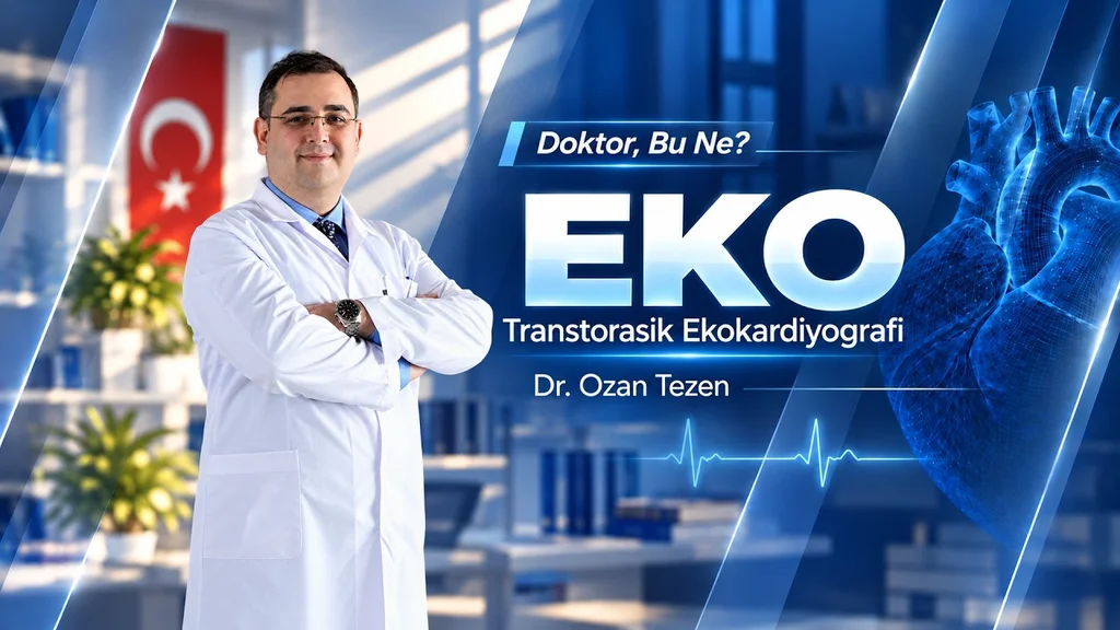
“Can you see my heart arteries during this test?” This is one of the questions our patients ask most often during an echocardiogram.
Echocardiography is a valuable way to assess the structure and function of the heart. Like every test, however, it has strengths and limitations. A routine echocardiogram is not a test designed to directly show narrowing in the coronary arteries.
What does it show? How do sound waves create images of the heart? Does it involve radiation? And how does an examination through the chest differ from one through the oesophagus? Let us take a closer look.
What Is Echocardiography?
Echocardiography uses ultrasound, or high-frequency sound waves, to create moving images of the heart. It is often called an “echo”.
During a standard examination, we assess two-dimensional views from different angles. These show the heart chambers, wall motion and valve opening and closing in real time. Suitable equipment and techniques can also provide three-dimensional images.
The basic principle is the same as ultrasound used during pregnancy or to examine abdominal organs. The transducer, or probe, and the characteristics of the sound waves vary according to the organ being examined.
How Is an Echocardiogram Performed?
An “echo” usually means transthoracic echocardiography: an examination performed through the chest wall.
Gel is applied and a probe is placed at particular points on the chest. Different positions provide different views. You may be asked to turn onto your side or briefly hold your breath to improve the images.
We watch the heart beating on the screen. The images allow us to measure the chambers and parts of the aorta, and assess how the heart contracts and how its valves work.
A standard chest echo does not require a catheter to be passed into an artery or the chest to be opened. It usually needs no special preparation, but instructions differ for other types of echo, so follow the advice provided when your appointment is arranged.
Does an Echocardiogram Involve Radiation?
No. Echocardiography uses sound waves, not X-rays or ionising radiation.
This allows the heart to be reassessed when clinically needed. Standard transthoracic echo is generally well tolerated, although pressure from the probe can cause mild discomfort.
An examination through the oesophagus is a different procedure, with its own preparation requirements and risks. Different types of echo should not be treated as identical examinations.
What Can an Echocardiogram Show?
Echo primarily provides information about the heart's structure and function. It can assess chamber size, wall thickness, pumping function and the movement of different regions of the heart wall.
The valves are examined for their structure, opening and closing, narrowing or leakage. Echo also plays an important role in assessing and monitoring heart failure.
Suitable views can help assess certain congenital heart problems and openings between the heart's chambers. However, not every opening or structural abnormality can be seen equally well with a standard examination.
The initial portions of the aorta near the heart can also be assessed. This does not mean that the entire aorta or the coronary arteries supplying the heart have been imaged in detail.
What Does Colour Doppler Do?
The colours on the echo screen come from colour Doppler imaging, which helps assess the direction and characteristics of blood flow.
When a valve leaks, backward flow can be assessed. Other Doppler measurements estimate blood-flow velocity and allow calculations relating to valve narrowing and certain pressure differences.
Colour alone is not a diagnosis. Images, measurements and the patient's clinical condition must be interpreted together. A colour change does not automatically mean an artery is blocked or a valve is leaking.
Can Echo Show the Coronary Arteries?
When patients refer to their “heart arteries”, they usually mean the coronary arteries supplying the heart muscle. Standard transthoracic echo does not assess their entire course and narrowing in the way angiography does.
In certain circumstances, the origins of coronary arteries or their flow can be assessed, but routine echo is not intended to map the coronary arteries.
Abnormal movement of the heart wall can provide clues to coronary artery disease even when the arteries are not assessed directly. However, these findings alone do not establish which artery is narrowed or by how much.
A normal echocardiogram does not conclusively exclude coronary artery narrowing. When direct imaging is needed, coronary CT angiography or invasive coronary angiography may be considered according to the clinical situation. This does not mean that every patient needs these tests.
Why Does Image Quality Vary?
During a chest examination, sound waves must travel to the heart and return. The chest wall, lungs and distance between the probe and the structure being examined can affect image quality.
Not every structure is equally clear in every patient. Some structures towards the back of the heart, particular openings or suspected blood clots may not be adequately assessed with standard imaging.
A structure being difficult to see does not necessarily mean it is abnormal. The clinician decides whether further imaging is needed based on the question being investigated.
Why Perform Echo Through the Oesophagus?
In transoesophageal echocardiography, a probe is passed through the mouth into the oesophagus. Because the oesophagus lies close to the back of the heart, certain structures can be examined in more detail from this position.
This may be needed for selected valve problems, artificial valves, suspected clots in particular areas or certain structural abnormalities. It is not required for everyone having an echo.
Both methods use sound waves; the main difference is where the images are obtained. The procedure, preparation and potential risks of transoesophageal echo are assessed and explained separately before it is performed.
Frequently Asked Questions About Echo
Are echo and ECG the same test?
No. An ECG records the heart's electrical activity. Echo uses sound waves to image its structure and movement. They answer different questions rather than replacing each other.
Does a normal echo rule out all heart disease?
No. A normal result provides valuable information about the structures and functions assessed, but does not exclude every heart condition, rhythm disorder or coronary artery narrowing. It must be considered alongside symptoms, examination findings and other tests.
Does a leaking valve on echo always require surgery?
No. The severity of leakage, valve structure, effects on the heart and symptoms are considered together. Treatment is not decided from a single phrase in an echo report.
Will I be asleep during an echo?
You are usually awake during a standard chest echo and can communicate with the person performing it. An examination through the oesophagus may require different preparation and sedation.
Conclusion
Echocardiography uses sound waves to assess the heart's structure, contraction and valves. It involves no ionising radiation and is used in the evaluation of many heart conditions.
However, it cannot answer every question on its own. Direct imaging of the coronary arteries and assessment of the heart's structure and function serve different purposes.
An echo result is best understood alongside your symptoms and other clinical findings.